45 muscle fiber model with labels
Solved art A rag the labels onto the diagram to identify the - Chegg Science. Anatomy and Physiology. Anatomy and Physiology questions and answers. art A rag the labels onto the diagram to identify the parts of a cardiac muscle fiber Reset Help tubule of DELL. Muscle Fiber Model (Altay) Flashcards | Quizlet Muscle fiber model identifications Terms in this set (21) sarcolemma Identify the membrane. endomysium Identify the tissue layer. myofibril Identify the structure. thick myofilament Identify the structure. thin myofilament Identify the structure. neuromuscular junction Identify the connection. axon Identify the structure. axon terminals
Muscle Fiber Model #1 - Ohio University - Anatomy & Physiology Muscle Fiber Model #1 - Ohio University - Anatomy & Physiology
Muscle fiber model with labels
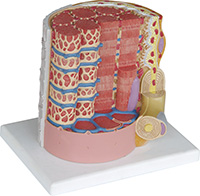
Sarcomere Model | Muscle Fiber Model | Skeletal Muscle | MICROanatomy ... This micro-anatomy model magnifies the anatomy of the human muscle fiber approximately 10,000 times. This muscle model illustrates a section of a skeletal muscle fiber and its neuromuscular end plate. The muscle fiber is the basic element of the diagonally striped skeletal muscle. You've never seen a muscle fiber in this way! Solved Cardiac muscle tissue: Identify and label- • | Chegg.com Anatomy and Physiology. Anatomy and Physiology questions and answers. Cardiac muscle tissue: Identify and label- • Striations Intercalated disc Nucleus • Branching • Cylindrical fiber . Quiz: Fill in the blank The heart has the following chambers: . . . Left ventricle sends blood to the right atrium receives blood from the Right atrium ... Muscles Labeling - The Biology Corner The activity linked below is a drag and drop activity for students to practice labeling the muscles, there are 6 slides showing images of muscles and fibers and the connective tissue surrounding the fibers (endomysium, perimysium, epimysium). Google Slides Key (TpT) Prev Article Next Article
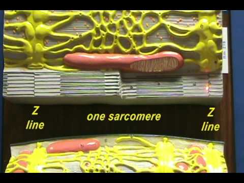
Muscle fiber model with labels. Skeletal Muscle Fiber Model - Myofibrils - YouTube This video was produced to help students of human anatomy at Modesto Junior College study our anatomical models. Muscle Fiber. 1. Myofibrils 2. Mitochondrium 3 ... - Pinterest Apr 24, 2014 - Muscle Fiber. 1. Myofibrils 2. Mitochondrium 3. Postsynaptic membrane 4. Synaptic gap with basal lamina 5. Presynaptic membrane 6. Presynaptic vesicle 7. Schwann cell 8. Nucleus 9. Actin filament 10. Sarcomere 11. Myosin filament 12. Myelin sheath 13. Neurofibers 14. Cell membrane (sarcolemma) 15. Transverse membrane tube 16. Triad 17. Sarcoplasmic reticulum 18. Basal lamina 19 ... Muscle Fiber Model: Motor Neuron, Myeline Sheath, Node of Ranvier ... Dec 20, 2013 - Muscle Fiber Model: Motor Neuron, Myeline Sheath, Node of Ranvier, Synaptic Terminal, Synaptic Cleft, Endomysium, Sarcolemma, Nuclei, Mitochondria, T ... General Anatomy of Skeletal Muscle Fibers - GetBodySmart Skeletal Muscle Fiber Location and Arrangement. are located inside muscles, where they are organized into bundles called […] Internal Anatomy of Skeletal Muscle Fibers. An interactive quiz about the internal anatomy of skeletal muscle fibers, featuring illustrations-based multiple choice questions.
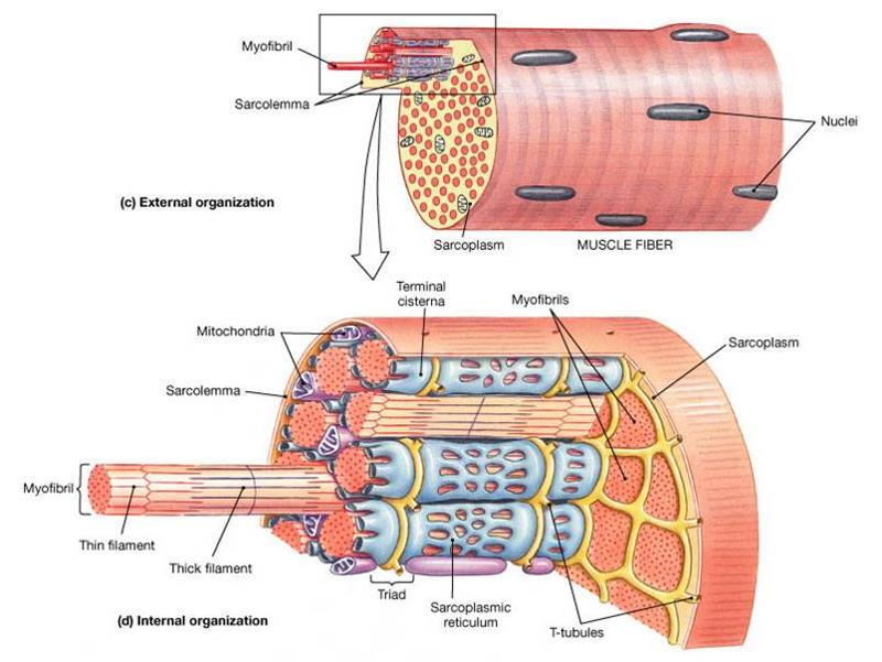
A 3D culture model of innervated human skeletal muscle enables studies ... 3D culture enhances skeletal muscle fiber maturation over 2D culture. (A) Representative confocal images of muscle fibers established in 2D (top row) and 3D conditions and immunostained for sarcomeric α-actinin (SAA; red), α-bungarotoxin (BTX; green), and Hoechst 33342 (blue) after 1, 2, and 3 weeks of culture.Scale bar, 50 μm. White arrowheads indicate broken fibers. Labeled Sarcomere Diagram The thin filaments Look at the diagram above and realize what happens as a muscle contracts. As will soon be described, the functional unit of a skeletal muscle fiber is the sarcomere, a highly organized arrangement of the contractile myofilaments actin .Play this quiz called Label the Sarcomere and show off your skills. Muscle Fiber Model Flashcards | Quizlet Muscle Fiber Model STUDY Flashcards Learn Write Spell Test PLAY Match Gravity Created by dee718 Key Concepts: Terms in this set (23) Myofibrils 1 Mitochondria 2 postsynaptic membrane 3 Synaptic gap with basal lamina 4 presynaptic membrane 5 presynaptic vesicles 6 satellite cell 7 Nucleus 8 myosin and actin filaments 9 Sarcomere 10 actin filaments 10.2 Skeletal Muscle - Anatomy & Physiology Figure 10.2.2 - Muscle Fiber: A skeletal muscle fiber is surrounded by a plasma membrane called the sarcolemma, which contains sarcoplasm, the cytoplasm of muscle cells. A muscle fiber is composed of many myofibrils, which contain sarcomeres with light and dark regions that give the cell its striated appearance. The Sarcomere
PDF THE MUSCULAR SYSTEM - University of Cincinnati Muscle fibers are organized into bundles supplied by blood vessels and innervated by motor neurons. Muscle structure. Skeletal (striated or voluntary) muscle consists of densely packed groups of hugely elongated cells known as myofibers. These are grouped into bundles (fascicles). Muscle fiber model Quiz - PurposeGames.com This is an online quiz called Muscle fiber model There is a printable worksheet available for download here so you can take the quiz with pen and paper. Your Skills & Rank Total Points 0 Get started! Today's Rank -- 0 Today 's Points One of us! Game Points 13 You need to get 100% to score the 13 points available Actions Add to favorites 5 favs SAC A&P Model Key - Muscular System - austincc.edu Muscular System. M1 - Muscled Arm. M2 - Muscle Leg. M3 - Female Muscle Figure. M4 - Microanatomy Muscle Fiber. M5 - Muscle Figure. Sarcomere Diagram Labeled - Wiring Diagram Pictures A myofibril (also known as a muscle fibril) is a basic rod-like unit of a muscle cell. Muscles are composed of tubular cells called myocytes, known as muscle fibers in striated muscle, and these cells in turn contain many chains of schematron.org are created during embryonic development in a process known as myogenesis..
Sarcomere Model Sarcomere Structure - YouTube
Altay Size: 110x19x14,5 Weight: approx. 5100 g. Full Description. Muscular Body - cod:6000.58. This 1/4 life-size model is a useful tool to study human superficial musculature. Significant structures are numbered and referenced on the accompanying k-card. Size: 25x18x40 cm Weight: 765 g. Full Description. Skeletal Muscle Fiber - cod:6000.32. This ...

Ben Smith: Textures (feels good)
Muscle Fiber Labeling Quiz - PurposeGames.com About this Quiz This is an online quiz called Muscle Fiber Labeling Quiz There is a printable worksheet available for download here so you can take the quiz with pen and paper. Your Skills & Rank Total Points 0 Get started! Today's Rank -- 0 Today 's Points One of us! Game Points 17 You need to get 100% to score the 17 points available Actions
Print Anatomy Exam 2 flashcards | Easy Notecards
Cardiac Muscle Fiber Model - spectrum-scientifics.com This block model depicts a cardiac muscle fiber that has been greatly enlarged to represent significant anatomical features such as the intercalated discs. Also includes detailed representations of sarcolemma, T-tubules, myofilaments, nucleus, and mitochondria. Mounted on a base. Dimensions: 15.75" x 10.25" x 7.5".

FIGURE
PDF MUSCLE MODEL ACTIVITY GUIDE - Field Museum muscle model / The Machine inside: BioMechanics activity guide For stud E nt ACTIVITY - Exploring Muscle Size Most animals, including humans, are born with the exact number of muscle fibers they will have their entire lives. This means they cannot increase their density, but they can change the size of muscle fibers. Explore how fiber size ...

Sarcomere Model | Muscle Fiber Model | Skeletal Muscle | MICROanatomy Muscle Fiber
Muscle Fibers: Anatomy, Function, and More - Healthline Muscle fibers are single muscle cells. When grouped together, they work to generate movement of your body and internal organs. You have three types of muscle tissue: skeletal, smooth, and cardiac....

V Ling: Art Center Summer Show UPDATE
Muscle Models | Muscle Figures | Musculature Models 3B MICROanatomy™ Human Muscle Fiber Model, 10,000 times magnified - 3B Smart Anatomy. $ 339.00. Item: 1000213 [B60] This micro-anatomy model magnifies the anatomy of the human muscle fiber approximately 10,000 times. This muscle model illustrates a section of a skeletal muscle fiber and its neuromuscular end plate.

Figure 1114 - Body Function - 78 Steps Health Journal
Anatomy Model Keys - Anatomical Models and Keys - NEOMED Library at ... Northeast Ohio Medical University is an Equal Education and Employment Institution ADA Compliance | Title IX. NEOMED Library- 4209 St, OH-44, Rootstown, OH 44272 - "A Building" Second Floor. 330-325-6600. library@neomed.edu. Except where otherwise noted, content on the NEOMED LibGuides is licensed for reuse under a Creative Commons 3.0 Attribution-NonCommercial license (CC BY-NC)

V Ling: Art Center Summer Show UPDATE
PDF Anatomy & Physiology - Truckee Meadows Community College Subcutis (Hypodermis) 1. External Horny Layer (Stratum corneum) 1a. Clear Layer (Stratum lucidum) -(KS 3 only) 2. Internal Hornless Germinative Zone (Stratum germinativum) 2a. Granular Layer (Stratum granulosum) 2b. Prickle-cell Layer (Stratum spinosum) 2c. Cylindrical Layer (Stratum basale) 3. Papillae 4.

Muscle Fiber Type and Training by Muhammed Javed - bodyandstrength.com
Muscles Labeling - The Biology Corner The activity linked below is a drag and drop activity for students to practice labeling the muscles, there are 6 slides showing images of muscles and fibers and the connective tissue surrounding the fibers (endomysium, perimysium, epimysium). Google Slides Key (TpT) Prev Article Next Article

V Ling: 08.10
Solved Cardiac muscle tissue: Identify and label- • | Chegg.com Anatomy and Physiology. Anatomy and Physiology questions and answers. Cardiac muscle tissue: Identify and label- • Striations Intercalated disc Nucleus • Branching • Cylindrical fiber . Quiz: Fill in the blank The heart has the following chambers: . . . Left ventricle sends blood to the right atrium receives blood from the Right atrium ...

ANATOMY & PHYSIOLOGY I BIS 240: Muscle Fiber Model B
Sarcomere Model | Muscle Fiber Model | Skeletal Muscle | MICROanatomy ... This micro-anatomy model magnifies the anatomy of the human muscle fiber approximately 10,000 times. This muscle model illustrates a section of a skeletal muscle fiber and its neuromuscular end plate. The muscle fiber is the basic element of the diagonally striped skeletal muscle. You've never seen a muscle fiber in this way!

Altay Anatomical Model Skeletal Muscle Fibre

Gantungan kunci karet, Frosted, Fiberglass, Digital printing, Emboss, Kaca patri, Seni kaca ...

Muscle Fiber Types

BIOL 220: Internal organization of a muscle fiber
Types Muscle Tissue Skeletal Muscle Smooth Stock Vector 126845972 - Shutterstock
TD Models

Chpt 9 Muscle/Muscle Fibers flashcards | Quizlet
Post a Comment for "45 muscle fiber model with labels"